Overview
what it is and why it matters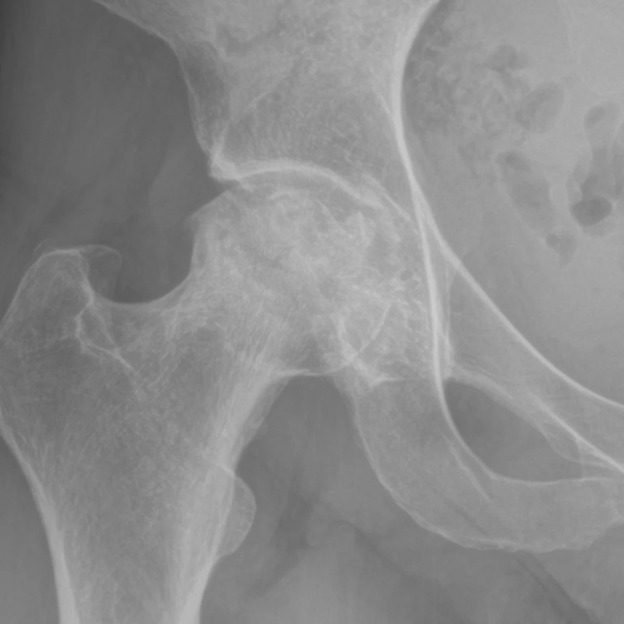
Avascular necrosis (AVN), also called osteonecrosis, is what happens when the blood supply to the ball at the top of your femur (the femoral head) is interrupted. Bone is living tissue that depends on a constant flow of blood to deliver oxygen and remove waste. The femoral head's supply is especially fragile: most of it enters through a narrow corridor along the neck of the bone, and any disruption, a fracture, a hip dislocation, or damage to the tiny vessels inside the bone itself, can cut off enough flow to kill a patch of bone.
Without blood, the dead bone cannot repair the microscopic fractures that normal daily loading creates. Those microfractures accumulate, the smooth surface of the ball begins to flatten and collapse, and once it collapses the cartilage on top crumbles with it, producing rapid and often severe arthritis of the hip joint. Common risk factors include high-dose or prolonged corticosteroid use (oral steroids for asthma, lupus, or transplant immunosuppression), excessive alcohol use, sickle cell disease, clotting disorders, and prior radiation. In many cases no clear cause is found (idiopathic AVN). The condition often affects both hips, in roughly half of cases, and it tends to strike adults in their 30s through 50s, earlier in life than typical wear-and-tear arthritis.
Symptoms
what patients describeEarly AVN is often silent, or the pain is so vague that it gets blamed on a muscle strain. The first symptom is usually a dull ache deep in the groin, not on the side of the hip or the buttock, but in the crease where your leg meets your trunk. The pain comes on with weight-bearing and is relieved by rest. Because the damage is inside the bone, the hip may still move normally at this stage, and a physical exam can be surprisingly unremarkable.
As the necrotic area enlarges, pain becomes more persistent. You may start to limp, and your provider may notice that the hip has lost some of its ability to rotate inward. A sudden worsening of pain, sometimes described as a sharp catch or a feeling that the hip "gave way," can signal that the femoral head has begun to collapse. Once collapse occurs, the progression to end-stage arthritis can be rapid, months rather than years.
Diagnosis
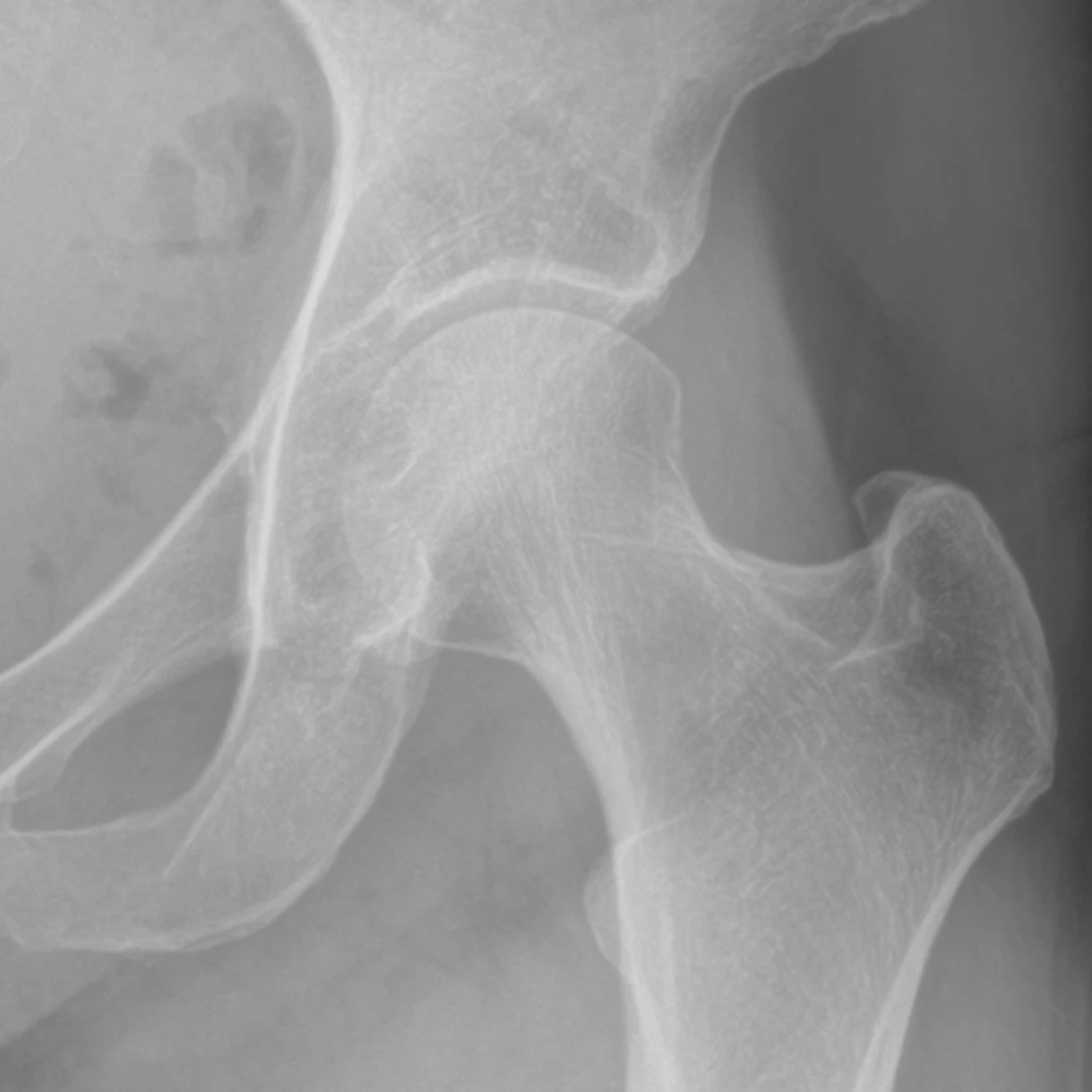
exam first, imaging secondX-rays of the hip are the first step, but they are often normal in early AVN, the bone has not yet changed shape. By the time the X-ray shows a thin bright line just under the surface of the femoral head, where the dead bone is separating from the living bone above it, or visible flattening of the ball, the disease is already advanced. That is why MRI is the gold-standard study: it can detect the dead-bone signal months before any X-ray change appears, when the window for preserving the joint is still open.
Once AVN is confirmed, your provider stages it based on how much of the femoral head is involved and whether the surface has started to collapse. Staging drives the treatment decision: early stages may respond to protective measures, while a collapsed femoral head almost always requires replacement. If one hip is affected, your provider often orders an MRI of the other hip as well, since the condition often affects both hips.
Treatment Path
how care progresses1
Protected Weight-Bearing
The idea is straightforward: if the bone is weakened but has not yet collapsed, reducing the load passing through it may buy time. Your provider may recommend crutches or a cane to limit how much force the femoral head absorbs with each step. Evidence that protected weight-bearing alone prevents collapse is limited, but it remains a reasonable first measure while further workup and staging are completed.
2
NSAIDs and Analgesics
Anti-inflammatory medication, ibuprofen, naproxen, or meloxicam, helps manage pain while your provider completes staging and discusses next steps. These medications do not change the course of the disease, but they can meaningfully improve day-to-day comfort during the evaluation window.
3
Bisphosphonates
Bisphosphonates are medications that slow the normal breakdown-and-rebuild cycle of bone. In early-stage AVN, the theory is that slowing bone resorption around the dead zone may help the femoral head hold its shape long enough for new blood vessels to grow in. Some studies show a delay in collapse; others show no benefit. Your provider weighs the stage, lesion size, and your overall risk profile before recommending a trial.
Surgical Options
if non-operative care isn't enoughMost patients with AVN beyond the earliest stages ultimately require surgery. The choice of procedure depends on the stage, the size of the necrotic lesion, and whether the femoral head has collapsed.
Frequently Asked
questions we hear in clinicWhy is the femoral head so prone to losing its blood supply?
Most of the blood supply to the femoral head enters through a narrow corridor along the neck of the bone. Because so much of it travels through that single tight pathway, a fracture, a hip dislocation, or damage to the tiny vessels inside the bone can cut off enough flow to kill a patch of bone.
What raises my risk of avascular necrosis?
Common risk factors include high-dose or prolonged corticosteroid use (oral steroids for asthma, lupus, or transplant immunosuppression), excessive alcohol use, sickle cell disease, clotting disorders, and prior radiation. In many cases no clear cause is found, which is called idiopathic AVN.
Why does early AVN often go unnoticed?
Early AVN is often silent, or the pain is so vague that it gets blamed on a muscle strain. The first symptom is usually a dull ache deep in the groin that comes on with weight-bearing and is relieved by rest. Because the damage is inside the bone, the hip may still move normally at this stage and a physical exam can be surprisingly unremarkable.
Do I need an MRI if my X-ray looks normal?
Often yes. X-rays are the first step but are frequently normal in early AVN, before the bone has changed shape. MRI is the gold-standard study because it can detect the dead-bone signal months before any X-ray change appears, when the window for preserving the joint is still open.
Will both of my hips be affected?
They might be. AVN often affects both hips, in roughly half of cases. If one hip is affected, your provider often orders an MRI of the other hip as well.
Can avascular necrosis be treated without surgery?
Early stages may respond to protective measures such as protected weight-bearing, anti-inflammatory medication for comfort, and in some cases bisphosphonates. However, most patients with AVN beyond the earliest stages ultimately require surgery, and a collapsed femoral head almost always requires replacement.
Providers Who Treat Avascular Necrosis of the Hip
sports-medicine team


Further Reading
authoritative sourcesExternal patient-education references and related OSI pages for additional background: