Overview
The tibia (tibia) is the main weight-bearing bone of the lower leg, and fractures of its shaft (the long middle portion) are among the most common serious leg injuries. They typically result from high-energy events such as motor-vehicle accidents, falls from height, or contact sports injuries. Because the tibia runs just beneath the skin along its front surface with little soft-tissue coverage, open fractures (ones where bone breaks through the skin) are more common here than in almost any other bone, and they require urgent treatment to reduce infection risk.
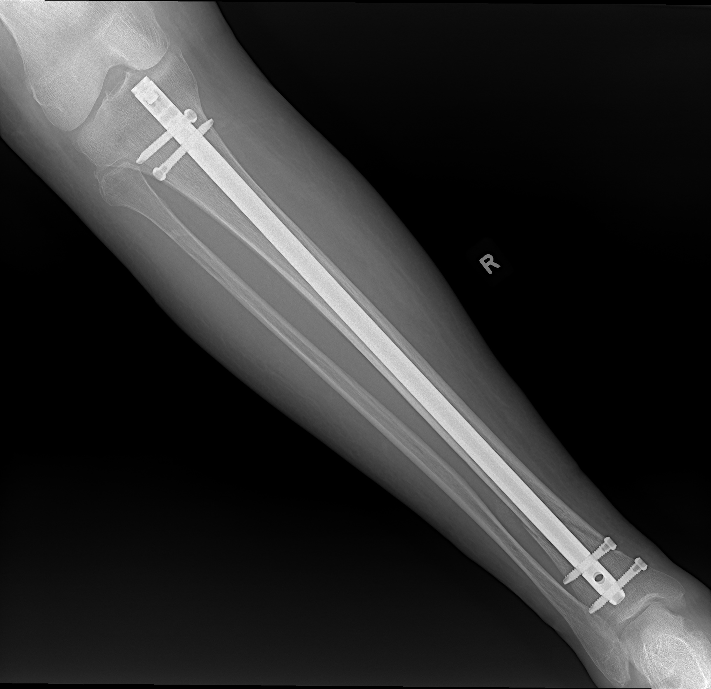
Intramedullary nailing (IMN) is the standard surgical treatment for displaced tibial shaft fractures. The procedure passes a metal rod down the hollow center of the bone, called the medullary canal, through small incisions far from the fracture itself. This approach restores the leg's length, alignment, and rotation without stripping away the soft-tissue envelope around the fracture, which preserves the blood supply and the biology needed for healing.
Unlike a cast, the nail holds the bone rigidly enough that most patients can put some weight on the leg within days of surgery. Early weight-bearing and motion reduce complications such as muscle wasting, blood clots, and joint stiffness, and they help patients return to daily activities sooner. The nail is designed to remain in place permanently, though a subset of patients later elect to have it removed if hardware-related knee pain develops.
The decision to operate depends on fracture displacement, stability, and the overall injury pattern. Fractures that can be held in a reliable position with a cast alone may not need surgery; a thorough evaluation, including X-rays and sometimes a CT scan, guides that decision.
Why it's done
Tibial shaft intramedullary nailing is typically considered when imaging and the clinical picture together indicate that the fracture will not reliably heal or function without surgical stabilization. Common indications include:
Displaced or unstable shaft fracture
Alignment cannot be reliably held in a cast.
Open fracture
Urgent irrigation, debridement, and stabilization.
Segmental fracture pattern
Two fracture levels are inherently unstable.
Polytrauma
Rapid stabilization aids mobilization and overall care.
Nonunion from prior non-operative care
Nailing plus bone grafting salvages most cases.
How it works
We make a small incision just above or below the kneecap and pass a guidewire down the hollow center of the tibia under live X-ray (fluoroscopy). The canal is then gently enlarged to accept the nail, which is driven down to bridge the fracture.
Interlocking screws above and below the fracture control rotation and length. Associated fibular fractures (the smaller bone in the lower leg) rarely need their own fixation. We confirm alignment, rotation, and nail position with fluoroscopy before closing.
Recovery
Most isolated tibial shaft fractures allow you to put weight on the leg shortly after surgery. Early knee and ankle motion begins immediately. We confirm healing on X-ray at follow-up visits. Knee pain at the nail entry site is relatively common after tibial nailing, and the nail can be removed once the bone has fully healed if that pain is bothersome. Slow healing and rotational malalignment are known complications.
Physicians Who Perform Tibial Shaft Intramedullary Nailing


Contact
For questions about this procedure or to schedule an evaluation, call the office at (830) 625-0009 or schedule an appointment online.
Risks & Why We Still Recommend It
Every operation carries risk. This procedure is offered because the condition, when left untreated, can cause a tibia that cannot bear weight and whose malalignment, once set, is hard to correct. The decision to proceed weighs the risks of surgery against the limitations the condition places on daily function. Surgery does not remove risk; it addresses a problem that is otherwise progressive. Whether it is appropriate is determined for each patient in consultation with the surgeon.
The risks we discuss with you before tibial shaft intramedullary nailing include:
- bleeding and infection
- anesthesia risk
- blood clot in the leg or lung
- compartment syndrome (the tibia is the most common site)
- anterior knee pain at the nail entry
- non-union or delayed union
- mal-union (particularly in proximal- or distal-third patterns)
- hardware irritation
The indication to proceed is a displaced or unstable tibial shaft fracture. If this operation is not right for you, we will not recommend it.
Frequently Asked
questions we hear in clinicWhy a rod instead of a cast?
Fractures that can be held in a reliable position with a cast alone may not need surgery. For displaced fractures, the nail restores the leg's length, alignment, and rotation without stripping away the soft-tissue envelope around the fracture, and unlike a cast it holds the bone rigidly enough that most patients can put some weight on the leg within days of surgery.
Will the incision be at the fracture site?
No. The rod passes down the hollow center of the bone through small incisions far from the fracture itself: a small incision just above or below the kneecap, with interlocking screws above and below the fracture to control rotation and length.
When can I walk on the leg?
Most isolated tibial shaft fractures allow you to put weight on the leg shortly after surgery, and early knee and ankle motion begins immediately. Early weight-bearing and motion reduce complications such as muscle wasting, blood clots, and joint stiffness.
Does the rod come out later?
The nail is designed to remain in place permanently. Knee pain at the nail entry site is relatively common after tibial nailing, and the nail can be removed once the bone has fully healed if that pain is bothersome.
What about the smaller bone in my lower leg?
Associated fibular fractures rarely need their own fixation.
What are the main risks?
Bleeding and infection, anesthesia risk, blood clot in the leg or lung, compartment syndrome (the tibia is the most common site), anterior knee pain at the nail entry, non-union or delayed union, mal-union, and hardware irritation.
Further Reading
External patient-education references and related OSI pages for additional background: